



The Process Behind Emergency Room X-ray Imaging Explained
2026-08-24


The Process Behind Emergency Room X-ray Imaging Explained
Introduction to X-ray Imaging in Emergency Rooms
In modern healthcare, **X-ray imaging** stands out as a vital tool, particularly in emergency room (ER) settings. This non-invasive diagnostic technique allows physicians to visualize the internal structure of the body, helping them quickly ascertain injuries or underlying health issues. Understanding the process behind **emergency room X-ray imaging** is crucial for patients and medical practitioners alike.
Understanding the Role of X-ray Imaging in Emergency Care
X-ray imaging plays a pivotal role in emergency medicine by facilitating rapid diagnosis. Conditions such as fractures, infections, and tumors can be identified swiftly, enabling healthcare providers to initiate appropriate treatment without delay. The efficiency of X-ray imaging directly impacts patient outcomes, making it a cornerstone of emergency care.
The Importance of Timely Diagnosis
In emergencies, every second counts. Delayed diagnoses can lead to complications and, in some cases, worsen the patient's condition. By utilizing X-ray technology, medical teams can expedite diagnosis and treatment, significantly improving survival rates and recovery times.
The X-ray Imaging Process in the Emergency Room
The X-ray process in an emergency room involves several key steps, from patient assessment to image analysis. Each phase is essential for ensuring high-quality results that contribute to effective treatment plans.
1. Patient Assessment and Preparation
Upon arrival at the emergency room, patients undergo a thorough assessment conducted by triage nurses or physicians. This evaluation includes medical history, symptoms, and physical examination. If an X-ray is warranted, the healthcare provider will explain the procedure to the patient, addressing any concerns and ensuring informed consent.
Preparing the Patient for X-ray Imaging
Patients may need to change into a hospital gown, depending on the area being examined. It's essential to remove any jewelry or clothing that could obstruct the imaging area to guarantee accurate results.
2. The X-ray Procedure
Once prepared, the patient is escorted to the X-ray room, where the imaging equipment is housed. The procedure is quick, typically lasting only a few minutes.
Positioning the Patient for Optimal Imaging
Proper positioning is critical to obtaining clear X-ray images. Radiologic technologists specialize in positioning patients to capture the required views while minimizing discomfort. They may employ various techniques, such as adjusting the X-ray machine's angle or utilizing supports for patients in pain.
Utilizing Advanced X-ray Technology
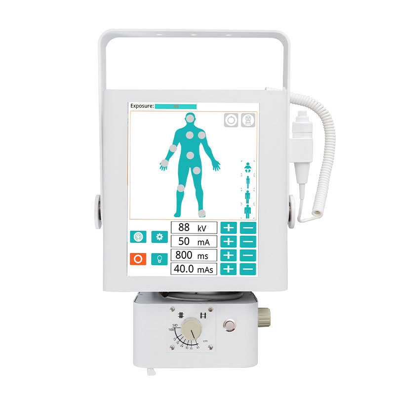
Modern X-ray machines are equipped with advanced features, including digital imaging and adjustable settings to enhance image quality. These technologies allow for faster processing times and more precise diagnostics, reducing the need for repeat imaging.
3. Image Acquisition and Processing
Once the patient is positioned, the technologist activates the X-ray machine. The machine emits controlled radiation that penetrates the body, creating images that are captured on a digital plate.
How Digital X-ray Imaging Works
Digital X-ray systems convert X-ray energy into electrical signals, which are processed to create images on a computer screen. This technology not only speeds up the imaging process but also allows for immediate review by healthcare providers.
4. Image Interpretation by Radiologists
After the images are obtained, they are reviewed by a radiologist, a medical doctor specialized in interpreting diagnostic images. The radiologist assesses the X-rays for any abnormalities, such as fractures, dislocations, or pathologies.
Reporting Results to the Emergency Team
The radiologist compiles their findings into a report, which is promptly communicated to the emergency care team. This swift exchange of information is crucial for determining the next steps in patient management and treatment.
Common Conditions Diagnosed Using X-ray Imaging
X-ray imaging is instrumental in diagnosing a range of conditions encountered in emergency settings. Some of the most common issues include:
1. Fractures and Dislocations
One of the primary uses of X-rays in the ER is identifying fractures and dislocations following traumatic injuries. X-rays can reveal the type and severity of the injury, guiding treatment options such as immobilization or surgical intervention.
2. Infections
X-rays can help identify infections in bones (osteomyelitis) or lungs (pneumonia). The images can show abnormalities in the affected areas, aiding physicians in selecting appropriate antibiotics or other treatments.
3. Tumors and Masses
In some cases, X-ray imaging can reveal the presence of tumors or unusual masses. This finding may warrant further investigations, including CT scans or biopsies, to determine the nature of the growth.
Safety and Risks Associated with X-ray Imaging
While X-ray imaging is generally safe, it’s essential to understand the associated risks and safety measures taken in emergency rooms.
Radiation Exposure Concerns
X-rays involve exposure to low levels of ionizing radiation. However, the benefits of accurate diagnosis often outweigh the risks. Emergency rooms follow strict guidelines to minimize exposure, ensuring that only necessary images are taken.
Protective Measures for Patients
To safeguard patients, healthcare teams utilize lead aprons and shields when possible. Additionally, protocols dictate the lowest radiation dose necessary to achieve quality images, effectively managing exposure.
Advancements in X-ray Technology
The field of radiology is continuously evolving, with advancements enhancing the efficacy of X-ray imaging in emergency care.
Digital Imaging Innovations
The transition from traditional film to digital X-rays has revolutionized diagnostic capabilities. Digital imaging allows for better image storage, retrieval, and sharing, improving collaboration among healthcare teams.
3D Imaging and Tomosynthesis
Recent innovations include 3D imaging and tomosynthesis. These techniques provide a more comprehensive view of structures, enhancing accuracy in diagnosing complex conditions, particularly in breast imaging.
The Role of Healthcare Professionals in X-ray Imaging
Effective X-ray imaging in emergency departments relies heavily on the expertise of various healthcare professionals.
Radiologic Technologists
Radiologic technologists play a critical role in operating X-ray equipment and ensuring patient comfort during the procedure. Their expertise is essential for obtaining high-quality images.
Radiologists
Radiologists are responsible for interpreting the X-ray images and providing vital insights to the emergency care team. Their specialized training equips them to identify nuances that might be missed by non-specialists.
Frequently Asked Questions (FAQs)
1. How long does an X-ray take in the emergency room?
The actual X-ray process typically takes just a few minutes. However, total time may vary based on patient volume and the need for additional imaging.
2. Is X-ray imaging safe for pregnant women?
X-ray imaging poses risks due to radiation exposure. In cases of potential pregnancy, healthcare providers evaluate the necessity of the X-ray and may recommend alternatives if appropriate.
3. Can I eat or drink before an X-ray?
Generally, patients can eat and drink unless instructed otherwise. However, if the X-ray involves the abdomen or gastrointestinal tract, fasting may be required.
4. How do I prepare for an X-ray in the emergency room?
Wear comfortable clothing, and be prepared to change into a gown. Remove any metal objects, including jewelry, that may interfere with the imaging process.
5. What happens if the X-ray shows a problem?
If the X-ray reveals abnormalities, the healthcare team will discuss the findings and recommend further testing or treatment options based on the diagnosis.
Conclusion
Understanding the process behind emergency room X-ray imaging highlights its significance in providing timely and accurate diagnoses. From patient assessment to image interpretation, each phase is crucial for delivering effective care. As technology advances, X-ray imaging continues to evolve, ensuring that emergency departments remain equipped to meet the demands of patient care. By leveraging the expertise of healthcare professionals and utilizing cutting-edge technology, we can achieve better outcomes for patients in urgent medical situations.
TAG:
Related Posts
Understanding the Role of X-ray Machines in Emergency Rooms




Address
No. 16, Phase IV, International Leather City, Xinji City, Hebei Province, China